 Chemist Emmanuel Delamarche held a thin slice of human thyroid tissue on a glass slide between his fingers. The tissue poses a mystery: does it contain a tumor or not? Delamarche, who works at IBM Research in Zurich, Switzerland, turned the slide around in his hand as he explained that the normal method of diagnosing a tumor involves splashing a chemical reagent, some of which are expensive, onto the uneven surface of the tissue and watching for it to react with disease markers. A pathologist “looks at them under a microscope, and he’s using his expertise, his judgment, and looks at what chemical he used, what type of color he can see and what part and he has to come up with a diagnosis,” Delamarche says, “he has a very, very hard job, OK?”
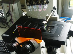
IBM is already good at precise application of materials to flat surfaces such as computer chips. Human tissue, sliced thin enough, turns out to receptive to the company’s bag of tricks too. Delamarche, turning to one of three machines on lab benches, explained that a few years ago his team began trying to deliver reagents with more precision. University Hospital Zurich will be testing the results over the next few months.
Read the rest of this post at IEEE Spectrum’s Tech Talk blog: [html] [pdf]